
Interesting Case March 2024
Case contributed by:
Ujunwa Korie, MD, MS and Yuanxin Liang, MD, PhD, Department of Pathology at Yale School of Medicine
Case History
A 78-year-old male presented with progressive dyspnea on exertion, lacking chest pains and
experiencing fatigue. His medical history included hypertension, hyperlipidemia, aortic valve
replacement, and chronic kidney disease stage IIIA. As a former smoker, he had quit 24 years
ago after a 30-pack year history. Workup revealed iron deficiency, and subsequent colonoscopy
exposed an ascending colon mass. Biopsy results indicated moderately differentiated
adenocarcinoma. Further CT imaging for staging revealed a 2 cm lesion in segment 4 of the left
hepatic lobe, raising suspicion of metastatic disease. A CT-guided percutaneous liver nodule
biopsy was subsequently performed.
Pathologic Findings
The liver biopsy exhibited well-preserved liver parenchyma with no notable abnormalities
(Figure 1). Additionally, a ciliated epithelial cyst lining surrounded by loose lamina propria,
smooth muscle, and an outer inconspicuous fibrous capsule was identified (Figures 2–3).
Notably, the ciliated cystic lining epithelial cells were negative for HNF1beta (Figure 4),
indicating non-biliary differentiation.
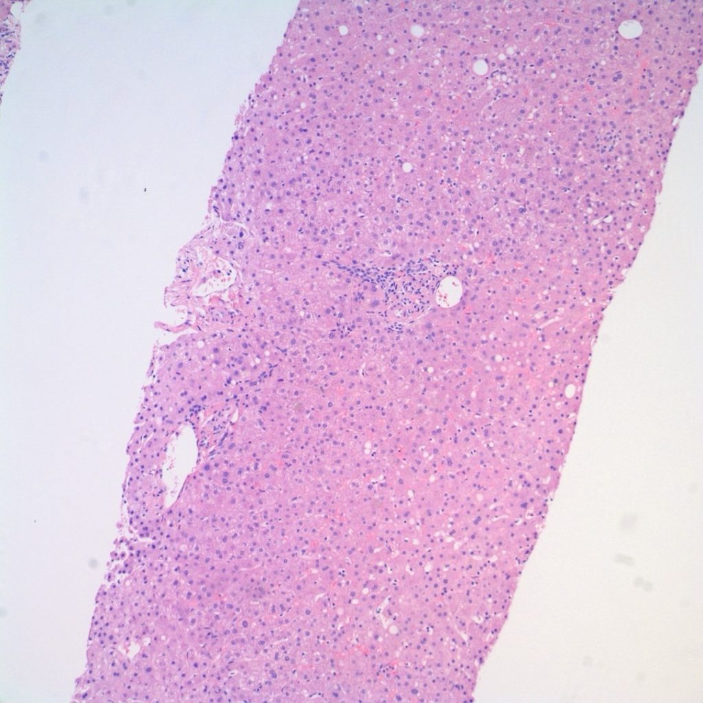
Figure 1. H&E slide demonstrates essentially normal liver parenchyma with portal tracts and lobules.
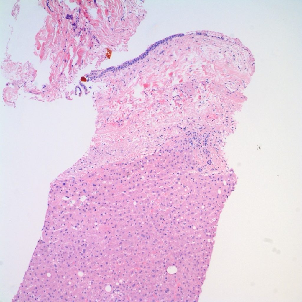
Figure 2: H&E low power image shows epithelial cyst lining with loose lamina propria and a smooth muscle layer.
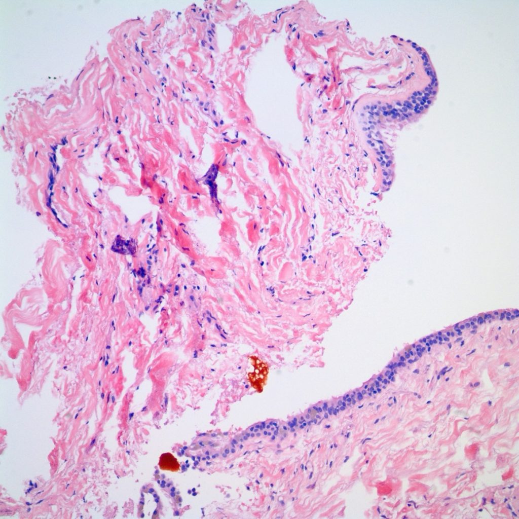
Figure 3a: H&E high power image demonstrates cilia.
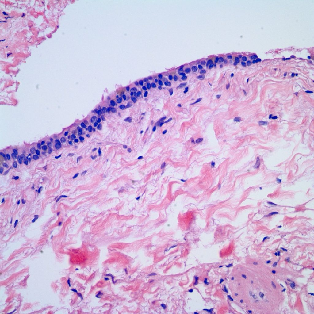
Figure 3b: H&E high power image demonstrates cilia.
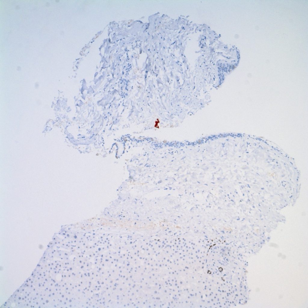
Figure 4: The immunostaining for HNF1beta highlights bile ducts in portal tract, while the cyst epithelial lining is negative for HNF1beta expression.
Diagnosis
Ciliated hepatic foregut cyst
Discussion
The ciliated hepatic foregut cyst is a rare cystic lesion originating from the embryological
foregut. The primitive foregut gives rise to the tracheobronchial tree, esophagus, and liver during
development. The liver specifically develops from the ventral foregut, while the tracheobronchial
tree and esophagus develop from the dorsal foregut. Bronchogenic cysts are distinguished by
ciliated columnar epithelium containing cartilage, smooth muscle, and mucous glands. In
contrast, esophageal cysts feature two smooth muscle layers and may exhibit either squamous or
columnar epithelium, which may be ciliated. (1)
The shared characteristics between ciliated hepatic foregut cyst (CHFC), bronchial and
esophageal cysts are believed to stem from the common embryological origin of the liver,
esophagus, and trachea, all of which develop from the foregut. The presence of these cysts is
thought to result from anomalous budding of foregut structures during the developmental
process. (1,3)
Grossly, the cyst is described as tan-pink solitary cyst, mostly unilocular with viscous, mucoid or
bloody contents. (8) Histologically, it is defined by the following characteristics:
(a) An inner layer of ciliated pseudostratified cuboidal to columnar epithelium (may have
goblet cells)
(b) Loose lamina propria
(c) One to three smooth muscle layers
(d) An outer fibrous capsule
These cysts are typically discovered incidentally through imaging, autopsy, or surgery (8).
Described as benign and solitary, they are generally small, measuring less than 4 cm. Their
predominant location is in segment 4 of the left lobe of the liver, although occurrences in the
right lobe have also been observed. There is a slightly higher incidence in men (9). Ciliated
hepatic foregut cyst is the only recognized primary hepatic entity with ciliated characteristics.
However, it is important to differentiate this cyst from other hepatic lesions, such as parasitic
cysts, simple hepatic cysts (also known as solitary bile duct cyst or benign hepatic cyst), and
hepatobiliary cystadenoma, as these lesions may potentially be malignant (1).
Although ciliated hepatic foregut cyst (CHFC) is generally regarded as benign, there have been
documented instances of squamous metaplasia and subsequent malignant transformation,
typically into squamous cell carcinoma. This highlights the importance of thorough sampling or
complete excision, especially in symptomatic cases of CHFC. Close clinical monitoring of
patients is advised in order to promptly detect any potential developments (4-7).
References
- Vick, Dan J. M.D.; Goodman, Zachary D. M.D., Ph.D.; Deavers, Michael T. M.D.; Cain, Joyce
B.S.; Ishak, Kamal G. M.D., Ph.D.. Ciliated Hepatic Foregut Cyst: A Study of Six Cases and
Review of the Literature. The American Journal of Surgical Pathology 23(6):p 671-677, June
1999. - Yang JD, Moon WS. Ciliated hepatic foregut cyst. Korean J Hepatol. 2012 Mar;18(1):98-100.
doi: 10.3350/kjhep.2012.18.1.98. Epub 2012 Mar 22. PMID: 22511910; PMCID: PMC3327000. - Terada, Tadashi M.D.; Nakanuma, Yasuni M.D.; Kono, Naoko Ph.D.; Ueda, Kazuhiko M.D.;
Kadoya, Masumi M.D.; Matsui, Osamu M.D.. Ciliated Hepatic Foregut Cyst: A Mucus
Histochemical Immunohistochenucal, and Ultrastructural Study in Three Cases in Comparison
with Normal Bronchi and Intrahepatic Bile Ducts. The American Journal of Surgical Pathology
14(4):p 356-363, April 1990. - Furlanetto, A., Dei Tos, A. Squamous cell carcinoma arising in a ciliated hepatic foregut
cyst. Virchows Arch 441, 296–298 (2002). https://doi.org/10.1007/s00428-002-0668-z - Anne-Sophie de Lajarte-Thirouard, Nathalie Rioux-Leclercq, Karim Boudjema, Yves Gandon,
Marie-Paule Ramée, Bruno Turlin, Squamous Cell Carcinoma Arising in a Hepatic Forgut Cyst,
Pathology – Research and Practice, Volume 198, Issue 10 - Ben Mena, N., Zalinski, S., Svrcek, M. et al. Ciliated hepatic foregut cyst with extensive
squamous metaplasia: report of a case. Virchows Arch 449, 730–733 (2006).
https://doi.org/10.1007/s00428-006-0320-4 - Xiangsheng Zhang, Zhen Wang, Yanjun Dong, Squamous cell carcinoma arising in a ciliated
hepatic foregut cyst: Case report and literature review, Pathology – Research and Practice, Volume
205, Issue 7, 2009, Pages 498-501, ISSN 0344-0338, https://doi.org/10.1016/j.prp.2008.12.003. - Joseph D. Jakowski, Joel G. Lucas, Sumit Seth, Wendy L. Frankel, Ciliated hepatic foregut cyst:
A rare but increasingly reported liver cyst, Annals of Diagnostic Pathology, Volume 8, Issue 6,
2004, Pages 342-346, ISSN 1092-9134, https://doi.org/10.1053/j.anndiagpath.2004.08.004 - Bishop, K.C., Perrino, C.M., Ruzinova, M.B. et al. Ciliated hepatic foregut cyst: a report of 6
cases and a review of the English literature. Diagn Pathol 10, 81 (2015).
https://doi.org/10.1186/s13000-015-0321-1